WWW.SCIENTIFICAMERICAN.COM
Biggest Brain Map Ever Shows Mouse Neurons in Stunning Detail
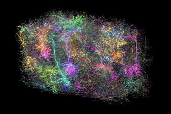
April 10, 20254 min readBiggest Brain Map Ever Shows Mouse Neurons in Stunning DetailNeuroscientists have created the largest and most detailed map of a mammal’s brain in a landmark achievementBy Miryam Naddaf & Nature magazine A rendering of more than 1,000 brain cells out of the those reconstructed from analysis of a cubic millimetre of brain tissue from a mouse. Allen InstituteResearchers have created the largest and most detailed wiring diagram of a mammalian brain to date, by mapping cells in a cubic millimetre of a mouse’s brain tissue. In a landmark achievement, the diagram also details the activity of individual neurons on a large scale―a neuroscience first.The high-resolution 3D map contains more than 200,000 brain cells, around 82,000 of which are neurons. It also includes more than 500 million of the neuronal connection points called synapses and more than 4 kilometres of neuronal wiring, all found in a tiny block of tissue in a brain region involved in vision. The only brain map of comparable scale is that of a cubic millimetre of human brain, which included 16,000 neurons and 150 million synapses. The new map also captured the activity of tens of thousands of neurons firing signals and interacting with each other to process visual information.This brain-activity map, combined with the wiring diagram, marks a milestone in connectomics, a field that aims to show how brains process and organize information. Behind the massive efforts are more than 150 researchers in the Machine Intelligence from Cortical Networks (MICrONS) project, who described their work in a package of eight papers published today in Nature and Nature Methods. The MICrONS project has made its resources available for the neuroscience community online, and other teams are already exploring them in different studies.On supporting science journalismIf you're enjoying this article, consider supporting our award-winning journalism by subscribing. By purchasing a subscription you are helping to ensure the future of impactful stories about the discoveries and ideas shaping our world today.“They managed to do something that we haven’t done as a neuroscience community in basically all of our history, which is to be able to map the activity of neurons onto the wiring on a very large population of neurons,” says Mariela Petkova, a neuroscientist at Harvard University in Cambridge, Massachusetts, who is not involved with the project. “We have never seen it at this scale.”The data “are really stunningly beautiful,” says Forrest Collman, a neuroscientist at the Allen Institute for Brain Science in Seattle, Washington, who co-authored the studies. “Looking at it really gives you an awe about the sense of complexity in the brain that is very much akin to looking up at the stars of night.”Mouse in a matrixTo create the breakthrough map, researchers first recorded the firing of almost 76,000 neurons in the visual cortex of a mouse as the animal watched various videos, including clips from The Matrix, for two hours. Then they sliced up a cubic millimetre of the mouse’s brain into thousands of tissue slices, each about one four-hundredth the width of a human hair.Rendering of a layer 5 Martinotti cell (grey) reconstructed from a large-scale electron microscopy dataset. The output synapses (bright spots) are color-coded by the target cell type. Red dots are synapses made onto excitatory layer 2/3 pyramidal cells and cyan dots are onto intra-telencephalic-projecting excitatory neurons in layer 5.Clare Gamlin/Allen InstituteThe scientists imaged each slice and assembled the images into a 3D map. Finally, they used artificial intelligence and machine-learning algorithms to annotate the neurons, their branching projections and their synapses. The team also matched the neurons in the map with their recordings of brain cells in action.Moritz Helmstaedter, a neuroscientist at the Max Planck Institute for Brain Research in Frankfurt, Germany, says “the combination of function and structure at that scale” is unprecedented. It’s “a very impressive endeavour and success”.Fire together, wire togetherThe work yielded insights into the basic rules that shape neural circuits in the mouse brain. For example, the authors found that neurons in the cortex that respond to similar types of visual feature—such as certain shapes or directions of movement—often form more connections with one another, no matter how far apart they are, than they do with neurons that specialize in a different type of feature.The results add a new wrinkle to a long-held theory in neuroscience, says Collman—namely, that “neurons that fire together wire together.” Previous studies have tested this theory only in limited numbers of neurons and synapses. The current study shows that “there’s a diversity [to] how much this rule is applied across all the different components of cortex,” he adds.The MICrONS researchers hope that their data set can help to reveal various features and processes in the brain. “There are all sorts of cortical areas that we understand at different levels of detail and in different ways. And I think this is really only the beginning of relating structure and function,” says Clay Reid, a neurobiologist at the Allen Institute and a co-author of the MICrONS papers.Helmstaedter says researchers can use the wiring maps to study how the brain stores and recalls visual memories, such as “our recollection of the last birthday party or of our grandparents.” These are “the big open questions about the mammalian cortex that are still very fundamental,” he adds.The newly published map covers around 0.2% of the mouse’s brain, but the MICrONS team will be testing the technologies to map the animal’s entire brain, says Nuno Maçarico da Costa, a neuroanatomist at the Allen Institute and another co-author of the MICrONS papers.This article is reproduced with permission and was first published on April 9, 2025.
0 Comments
0 Shares
51 Views


