


Can 3D printed implants restore erectile function? New research shows promising results
3dprintingindustry.com
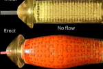
A group of Chinese researchers have developed a novel approach for addressing erectile dysfunction (ED) using biomedical 3D printing.In what is believed to be a world first, scientists successfully tested a 3D printed penile implant system in animals, reporting full restoration of erectile function in treated subjects. Published in Nature Biomedical Engineering, their findings offer promising insights into regenerative solutions for ED, a condition that affects more than 40% of men over 40, according to South China Morning Post.The study involved hydrogel-based bioinks to create an implant that closely mimics the anatomical and functional components of natural erectile tissue. Tested on pigs and rabbits, the technology yielded exceptional results while untreated subjects had a 25% reproductive success rate, those that received the implant showed a 100% success rate in mating and reproduction.Alongside South China University of Technology (SCUT), contributions also came from Guangzhou Medical University, Tokyo Medical and Dental University, and Columbia University.Lead author Wang Yingjun, an academician at the Chinese Academy of Engineering and President of the National Engineering Research Centre for Tissue Restoration and Reconstruction at SCUT, said, These findings indicate that the implants markedly improved functional recovery.The researchers used a hydrogel to 3D print a model of the corpus cavernosum a key structure in the penis that fills with blood during an erection. Next, they seeded this scaffold with endothelial cells the main cells that line blood vessels. Image via SCUT.A complex structure recreated with precisionNaturally, the penis has one of the most intricate vascular networks in the body, making reconstruction particularly challenging. Two corpus cavernosa run along its length, playing a key role in erections, while the tunica albuginea, a tough connective tissue layer, helps sustain them.To replicate these structures, researchers developed a hydrogel-based bioink, primarily composed of acrylic acid gelatin, to 3D print the corpora cavernosa. The implant was then encased in a fiber-based artificial tunica albuginea, providing the necessary strength to maintain function.For a more realistic and functional reconstruction, the team also 3D printed the corpus spongiosum, another erectile column, and the glans penis, assembling all components to mirror natural anatomy. To improve biocompatibility and reduce the risk of immune rejection, a layer of endothelial cells was added to the surface, supporting natural integration into the body.The study divided subjects into three groups: one received the 3D printed implant alone, another received both the implant and endothelial cells, while a control group with penile injuries received no treatment.The control group showed a 25% reproductive success rate, while those with 3D printed implants alone reached 75%. For the group that also received endothelial cells, the success rate climbed to 100%, indicating that the additional cell layer enhanced tissue regeneration and function.Recovery was swift. Two weeks after surgery, the animals regained normal erectile function, and within six weeks, they successfully mated and reproduced. The researchers noted that the findings suggest 3D printed hydrogel implants could restore damaged erectile tissue to near-normal function.Beyond ED treatment, the study highlights the potential of 3D printed functional tissue models for other organs with intricate circulatory networks, such as the heart and lungs. While previous research has explored these models, large-scale animal testing has been limited. The researchers emphasized that their study provides valuable insights into how 3D printed implants could translate into real-world applications, particularly in regenerative medicine.Although human trials are still a long way off, the study presents an important foundation for future research. If similar success is achieved in humans, this approach could lead to personalized, biologically compatible solutions for ED, offering an alternative to existing treatments.Local deformation to damage and flow measurement. Image via SCUT.3D printing for vascular organ reconstructionSCUTs approach aligns with broader efforts in bioprinting, where researchers are developing vascularized tissues, such as engineered blood vessels and functional heart models, to improve transplant success and advance regenerative medicine.For instance, Pohang University of Science and Technology (POSTECH), The Catholic University of Korea, and City University of Hong Kong (CityUHK) researchers successfully 3D printed biomimetic blood vessels and implanted them in a living rat, demonstrating a potential breakthrough in vascular grafts for cardiovascular disease treatment.Using a triple-coaxial cell printing technique and a specialized bioink made from smooth muscle and endothelial cells, the team developed functional vascular structures that integrated with living tissue over several weeks. The study suggested that these engineered blood vessels could offer a durable alternative for small-diameter vascular grafts, with future research focusing on enhancing their strength and evaluating long-term performance for human applications.In the US, researchers from the University of Minnesota developed a bio-ink that enabled them to 3D print a functional beating human heart, contributing a novel approach in cardiac tissue engineering.Leveraging pluripotent stem cells, they created an aortic replica with enhanced chamber structure and cell wall thickness, overcoming previous limitations in cardiac bioprinting. The printed heart maintained its electromechanical function for over six weeks, demonstrating potential applications in drug testing, disease modeling, and regenerative medicine.What3D printing trendsshould you watch out for in 2025?How is thefuture of 3D printingshaping up?To stay up to date with the latest 3D printing news, dont forget to subscribe to the 3D Printing Industry newsletter or follow us on Twitter, or like our page on Facebook.While youre here, why not subscribe to our Youtube channel? Featuring discussion, debriefs, video shorts, and webinar replays.Featured image shows the researchers used a hydrogel to 3D print a model of the corpus cavernosum a key structure in the penis that fills with blood during an erection. Next, they seeded this scaffold with endothelial cells the main cells that line blood vessels. Image via SCUT.
0 Comments
·0 Shares
·16 Views