Scientists figure out how the brain forms emotional connections
It's shocking!
Scientists figure out how the brain forms emotional connections
Neural recordings track how neurons link environments to emotional events.
Jacek Krywko
–
May 21, 2025 4:07 pm
|
16
Credit:
fotografixx
Credit:
fotografixx
Story text
Size
Small
Standard
Large
Width
*
Standard
Wide
Links
Standard
Orange
* Subscribers only
Learn more
Whenever something bad happens to us, brain systems responsible for mediating emotions kick in to prevent it from happening again. When we get stung by a wasp, the association between pain and wasps is encoded in the region of the brain called the amygdala, which connects simple stimuli with basic emotions.
But the brain does more than simple associations; it also encodes lots of other stimuli that are less directly connected with the harmful event—things like the place where we got stung or the wasps’ nest in a nearby tree. These are combined into complex emotional models of potentially threatening circumstances.
Till now, we didn’t know exactly how these models are built. But we’re beginning to understand how it’s done.
Emotional complexity
“Decades of work has revealed how simple forms of emotional learning occurs—how sensory stimuli are paired with aversive events,” says Joshua Johansen, a team director at the Neural Circuitry of Learning and Memory at RIKEN Center for Brain Science in Tokyo. But Johansen says that these decades didn’t bring much progress in treating psychiatric conditions like anxiety and trauma-related disorders. “We thought if we could get a handle of more complex emotional processes and understand their mechanisms, we may be able to provide relief for patients with conditions like that,” Johansen claims.
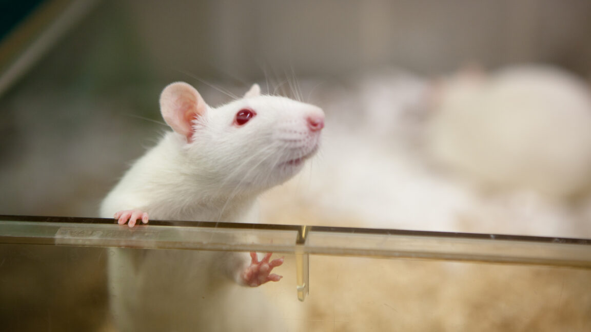
To make it happen, his team performed experiments designed to trigger complex emotional processes in rats while closely monitoring their brains.
Johansen and Xiaowei Gu, his co-author and colleague at RIKEN, started by dividing the rats into two groups. The first “paired” group of rats was conditioned to associate an image with a sound. The second “unpaired” group watched the same image and listened to the same sound, but not at the same time. This prevented the rats from making an association.
Then, one day later, the rats were shown the same image and treated with an electric shock until they learned to connect the image with pain. Finally, the team tested if the rats would freeze in fear in response to the sound. The “unpaired” group didn’t. The rats in the “paired” group did—it turned out human-like complex emotional models were present in rats as well.
Once Johansen and Gu confirmed the capacity was there, they got busy figuring out how it worked exactly.
Playing tag
“Behaviorally, we measured freezing responses to the directly paired stimulus, which was the image, and inferred stimulus which was the sound,” Johansen says. “But we also performed something we called miniscope calcium imaging.” The trick relied on injecting rats with a virus that forced their cells to produce proteins that fluoresce in response to increased levels of calcium in the cells. Increased levels of calcium are the telltale sign of activity in neurons, meaning the team could see in real time which neurons in rats’ brains lit up during the experiments.
It turned out that the region crucial for building these complex emotional models was not the amygdala, but the dorsomedial prefrontal cortex, which had a rather specialized role. “The dmPFC does not form the sensory model of the world. It only cares about things when they have emotional relevance,” Johansen explains. He said there wasn’t much change in neuronal activity during the sensory learning phase, when the animals were watching the image and listening to the sound. The neurons became significantly more active when the rats received the electric shock.
In the “unpaired” group, the active neurons that held the representations of the electric shock and the image started to overlap. In the “paired” group, this overlap also included the neuronal representation of the sound. “There was a kind of an associative bundle that formed,” Johansen says.
After Johansen and Gu pinpointed the neurons that formed those associative bundles, they started looking at how each of these components works.
Detraumatizing rodents
In the first step, the team identified the dmPFC neurons that sent output to the amygdala. Then they selectively inhibited those neurons and exposed the rats from the “paired” group to the image and the sound again. The result of disconnecting the dmPFC neurons from the amygdala was that rats exhibited a fear response to the image but no longer feared the sound. “It seems like the amygdala can form the simple representations on its own but requires input from the dmPFC to express more complex, inferred emotions,” Johansen says.
But there are still a lot of unanswered questions left.
The next thing the team wants to take a closer look at is the process that enables the brain to tie an aversive stimulus, like the shock, to one that was not active during the aversive event. In the “paired” group of rats, some multi-sensory neurons responding to both auditory and visual stimuli apparently got recruited. “We haven’t worked that out yet,” Johansen says. "This is a very novel type of mechanism.”
Another thing is that the emotional model Johansen and Gu induced in rats was relatively simple. In the real world, especially in humans, we can have many different aversive outcomes tied to the same triggers. A single location could be where you got stung by a wasp, attacked by a dog, robbed of your wallet, and dumped by your significant other—all different aversive representations with myriad inferred, indirect stimuli to go along with them. “Does the dmPFC combine all those representations into sort of a single, overlapping representation? Or is it a really rich environment that bundles different aversive experiences with the individual aspects of these experiences?” Johansen asked. “This is something we want to test more.”
Nature, 2025. DOI: 10.1038/s41586-025-09001-2
Jacek Krywko
Associate Writer
Jacek Krywko
Associate Writer
Jacek Krywko is a freelance science and technology writer who covers space exploration, artificial intelligence research, computer science, and all sorts of engineering wizardry.
16 Comments
#scientists #figure #out #how #brain
Scientists figure out how the brain forms emotional connections
It's shocking!
Scientists figure out how the brain forms emotional connections
Neural recordings track how neurons link environments to emotional events.
Jacek Krywko
–
May 21, 2025 4:07 pm
|
16
Credit:
fotografixx
Credit:
fotografixx
Story text
Size
Small
Standard
Large
Width
*
Standard
Wide
Links
Standard
Orange
* Subscribers only
Learn more
Whenever something bad happens to us, brain systems responsible for mediating emotions kick in to prevent it from happening again. When we get stung by a wasp, the association between pain and wasps is encoded in the region of the brain called the amygdala, which connects simple stimuli with basic emotions.
But the brain does more than simple associations; it also encodes lots of other stimuli that are less directly connected with the harmful event—things like the place where we got stung or the wasps’ nest in a nearby tree. These are combined into complex emotional models of potentially threatening circumstances.
Till now, we didn’t know exactly how these models are built. But we’re beginning to understand how it’s done.
Emotional complexity
“Decades of work has revealed how simple forms of emotional learning occurs—how sensory stimuli are paired with aversive events,” says Joshua Johansen, a team director at the Neural Circuitry of Learning and Memory at RIKEN Center for Brain Science in Tokyo. But Johansen says that these decades didn’t bring much progress in treating psychiatric conditions like anxiety and trauma-related disorders. “We thought if we could get a handle of more complex emotional processes and understand their mechanisms, we may be able to provide relief for patients with conditions like that,” Johansen claims.
To make it happen, his team performed experiments designed to trigger complex emotional processes in rats while closely monitoring their brains.
Johansen and Xiaowei Gu, his co-author and colleague at RIKEN, started by dividing the rats into two groups. The first “paired” group of rats was conditioned to associate an image with a sound. The second “unpaired” group watched the same image and listened to the same sound, but not at the same time. This prevented the rats from making an association.
Then, one day later, the rats were shown the same image and treated with an electric shock until they learned to connect the image with pain. Finally, the team tested if the rats would freeze in fear in response to the sound. The “unpaired” group didn’t. The rats in the “paired” group did—it turned out human-like complex emotional models were present in rats as well.
Once Johansen and Gu confirmed the capacity was there, they got busy figuring out how it worked exactly.
Playing tag
“Behaviorally, we measured freezing responses to the directly paired stimulus, which was the image, and inferred stimulus which was the sound,” Johansen says. “But we also performed something we called miniscope calcium imaging.” The trick relied on injecting rats with a virus that forced their cells to produce proteins that fluoresce in response to increased levels of calcium in the cells. Increased levels of calcium are the telltale sign of activity in neurons, meaning the team could see in real time which neurons in rats’ brains lit up during the experiments.
It turned out that the region crucial for building these complex emotional models was not the amygdala, but the dorsomedial prefrontal cortex, which had a rather specialized role. “The dmPFC does not form the sensory model of the world. It only cares about things when they have emotional relevance,” Johansen explains. He said there wasn’t much change in neuronal activity during the sensory learning phase, when the animals were watching the image and listening to the sound. The neurons became significantly more active when the rats received the electric shock.
In the “unpaired” group, the active neurons that held the representations of the electric shock and the image started to overlap. In the “paired” group, this overlap also included the neuronal representation of the sound. “There was a kind of an associative bundle that formed,” Johansen says.
After Johansen and Gu pinpointed the neurons that formed those associative bundles, they started looking at how each of these components works.
Detraumatizing rodents
In the first step, the team identified the dmPFC neurons that sent output to the amygdala. Then they selectively inhibited those neurons and exposed the rats from the “paired” group to the image and the sound again. The result of disconnecting the dmPFC neurons from the amygdala was that rats exhibited a fear response to the image but no longer feared the sound. “It seems like the amygdala can form the simple representations on its own but requires input from the dmPFC to express more complex, inferred emotions,” Johansen says.
But there are still a lot of unanswered questions left.
The next thing the team wants to take a closer look at is the process that enables the brain to tie an aversive stimulus, like the shock, to one that was not active during the aversive event. In the “paired” group of rats, some multi-sensory neurons responding to both auditory and visual stimuli apparently got recruited. “We haven’t worked that out yet,” Johansen says. "This is a very novel type of mechanism.”
Another thing is that the emotional model Johansen and Gu induced in rats was relatively simple. In the real world, especially in humans, we can have many different aversive outcomes tied to the same triggers. A single location could be where you got stung by a wasp, attacked by a dog, robbed of your wallet, and dumped by your significant other—all different aversive representations with myriad inferred, indirect stimuli to go along with them. “Does the dmPFC combine all those representations into sort of a single, overlapping representation? Or is it a really rich environment that bundles different aversive experiences with the individual aspects of these experiences?” Johansen asked. “This is something we want to test more.”
Nature, 2025. DOI: 10.1038/s41586-025-09001-2
Jacek Krywko
Associate Writer
Jacek Krywko
Associate Writer
Jacek Krywko is a freelance science and technology writer who covers space exploration, artificial intelligence research, computer science, and all sorts of engineering wizardry.
16 Comments
#scientists #figure #out #how #brain
0 Commentarii
·0 Distribuiri
·0 previzualizare


