A breakthrough 50 3D printed microscope by Strathclyde researchers
3dprintingindustry.com
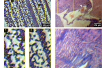
Researchers at the University of Strathclyde in Glasgow have created a fully 3D printed and low cost microscope for histological imaging.Based on OpenFlexures open-source design, the device incorporates 3D printed mechanical and optical components alongside a Raspberry Pi for control, an LED light source for illumination, and an affordable off-the-shelf camera. Weighing just 3 kg, the microscope can be assembled in three hours at an estimated cost of 50 ($60). This study is published in the bioRvix journal.As reported by New Scientist, Gail McConnell Professor of Biophotonics at the University, highlighted the impact of their work. She stated that her team had earlier devised a technique for 3D printing lenses commonly used in microscopes, a development that played a crucial role in achieving this milestone.Measuring the resolution of a fully 3D printed microscope post-acquisition chromatic correction. Image via University of Strathclyde.Affordability with 3D printed opticsKey to the projects success is the use of 3D printed lenses, fabricated using using an Elegoo Mars 3 Pro 3D printer and a photopolymerizing clear resin from Formlabs. Designed to replicate the specifications of traditional glass lenses, such as Edmund Optics 12.7 mm diameter plano-convex lens with a 35 mm focal length, these lenses achieve high performance at a fraction of the cost.The resins refractive index closely matches BK7 glass, commonly used in commercial optics. Through a meticulous post-processing workflow, the lenses were refined to meet the optical quality standards required for microscopy.Performance tests included analyzing a Giemsa-stained blood smear and a Haemotoxylin and Eosin-stained mouse kidney section. Images captured revealed intricate sub-cellular structures, including individual blood cells and renal tubules.With a magnification of 2.9x and a spatial resolution of approximately 5 micrometers, the microscope demonstrated capabilities on par with many commercial systems while maintaining a field of view measuring 1.7 mm.According to the researchers, relying on traditional glass lenses has long posed a challenge for cost-sensitive applications, especially in resource-limited settings. The integration of 3D printed optics addresses this issue, drastically reducing expenses while maintaining comparable imaging quality. They highlighted the accessibility of the device for low-budget laboratories, classrooms, and field diagnostics.The researchers also acknowledged certain limitations, such as uneven illumination across the field of view, resulting from alignment challenges with the LED light source. They proposed design modifications, including the addition of adjustable diaphragms, to improve illumination uniformity.Moreover, the customization potential of 3D printed lenses allows for the creation of alternative magnifications and numerical apertures, tailoring the device to specific applications.Having now demonstrated the feasibility of fabricating both mechanical and optical components through 3D printing, this work showcases a new approach to scientific instrumentation.(a) An image acquired of a Giemsa-stained blood smear. Two ROIs are shown, where individual red blood cells can be resolved over the 1.7 mm field of view. (b) An image of a H&E-stained mouse kidney. The thin section shows structures such as an interlobular arteriole (white arrow) and renal tubules, with a magnified ROI showing the organisation of nephrons in a medullary ray spanning the corticomedullary junction. Image via University of Strathclyde.Cost-effective 3D printed microscopesFurther showcasing the potential of 3D printed microscopes, University of Bath researchers developed an OpenFlexure 3D printed microscope, an open-source, laboratory-grade device that cost just $18 to produce.Designed for accessibility, the microscope featured motorized sample positioning, focus control, and a precise mechanical stage. Its low cost and customizable design aimed to democratize access to high-quality microscopy for schools, laboratories, clinics, and even homes.Initiated by Dr. Richard Bowman and maintained by Bath Open Instrumentation Group (BOING), this project ensured affordability without sacrificing imaging quality. As of 2020, over 100 units were produced in Tanzania and Kenya, where local adaptations highlighted the benefits of open-source designs. Co-creator Dr. Joel Collins emphasized its potential to advance education, research, and healthcare globally.One year before that, University of Connecticut and the University of Memphis researchers developed a 3D printed digital holographic microscopy (DHM) microscope capable of producing high-resolution 3D images at a low cost.Detailed in Optics Letters, the device provided twice the resolution of conventional DHM systems and eliminated the need for complex laboratory setups, making it suitable for field use in remote areas. Made entirely from 3D printed parts and common optical components, the microscope was compact, robust, and cost only a few hundred dollars to manufacture.By incorporating structured illumination (SI) technology, the microscope achieved a resolution of 0.775 m, allowing for precise imaging of biological samples like green algae. The team aimed to expand its use in biomedical diagnostics, particularly in underserved regions.Who won the 20243D Printing Industry Awards?All the news fromFormnext 2024.To stay up to date with the latest 3D printing news, dont forget to subscribe to the 3D Printing Industry newsletter or follow us on Twitter, or like our page on Facebook.While youre here, why not subscribe to our Youtube channel? Featuring discussion, debriefs, video shorts, and webinar replays.Featured image shows (a) An image acquired of a Giemsa-stained blood smear. Two ROIs are shown, where individual red blood cells can be resolved over the 1.7 mm field of view. (b) An image of a H&E-stained mouse kidney. The thin section shows structures such as an interlobular arteriole (white arrow) and renal tubules, with a magnified ROI showing the organisation of nephrons in a medullary ray spanning the corticomedullary junction. Image via University of Strathclyde.Ada ShaikhnagWith a background in journalism, Ada has a keen interest in frontier technology and its application in the wider world. Ada reports on aspects of 3D printing ranging from aerospace and automotive to medical and dental.
0 Yorumlar
·0 hisse senetleri
·38 Views


