How AM Elevates Healthcare: Insights from the Materialise 3D Printing in Hospitals Forum 2025
The cobbled streets and centuries-old university halls of Leuven recently served as a picturesque backdrop for the Materialise 3D Printing in Hospitals Forum 2025. Belgium’s Flemish Brabant capital hosted the annual meeting, which has become a key gathering for the medical 3D printing community since its launch in 2017.
This year, 140 international healthcare professionals convened for two days of talks, workshops, and lively discussion on how Materialise’s software enhances patient care. The Forum’s opening day, hosted at Leuven’s historic Irish College, featured 16 presentations by 18 healthcare clinicians and medical 3D printing experts.
While often described as the future of medicine, personalized healthcare has already become routine in many clinical settings. Speakers emphasized that 3D printing is no longer merely a “cool” innovation, but an essential tool that improves patient outcomes. “Personalized treatment is not just a vision for the future,” said Koen Peters, Executive Vice President Medical at Materialise. “It’s a reality we’re building together every day.”
During the forum, practitioners and clinical engineers demonstrated the critical role of Materialise’s software in medical workflows. Presentations highlighted value across a wide range of procedures, from brain tumour removal and organ transplantation to the separation of conjoined twins and maxillofacial implant surgeries. Several use cases demonstrated how 3D technology can reduce surgery times by up to four times, enhance patient recovery, and cut hospital costs by almost £6,000 per case.
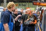
140 visitors attended the Materialise 3D Printing in Hospitals Forum 2025. Photo via Materialise.
Digital simulation and 3D printing slash operating times
Headquartered a few miles outside Leuven’s medieval center, Materialise is a global leader in medical 3D printing and digital planning. Its Mimics software suite automatically converts CT and MRI scans into detailed 3D models. Clinicians use these tools to prepare for procedures, analyse anatomy, and create patient-specific models that enhance surgical planning.
So far, Materialise software has supported more than 500,000 patients and analysed over 6 million medical scans. One case that generated notable interest among the Forum’s attendees was that of Lisa Ferrie and Jiten Parmar from Leeds General Infirmary. The pair worked alongside Asim Sheikh, a Consultant Skullbase and Neurovascular Neurosurgeon, to conduct the UK’s first “coach door osteotomy” on Ruvimbo Kaviya, a 40-year-old nurse from Leeds.
This novel keyhole surgery successfully removed a brain tumor from Kaviya’s cavernous sinus, a hard-to-reach area behind the eyes. Most surgeries of this kind require large incisions and the removal of substantial skull sections, resulting in extended recovery time and the risk of postoperative complications. Such an approach would have presented serious risks for removing Kaviya’s tumor, which “was in a complex area surrounded by a lot of nerves,” explained Parmar, a Consultant in Maxillofacial Surgery.
Instead, the Leeds-based team uses a minimally invasive technique that requires only a 1.5 cm incision near the side of Ravimbo’s eyelid. A small section of skull bone was then shifted sideways and backward, much like a coach door sliding open, to create an access point for tumor removal. Following the procedure, Ravimbo recovered in a matter of days and was left with only a 6 mm scar at the incision point.
Materialise software played a vital role in facilitating this novel procedure. Ferrie is a Biomedical Engineer and 3D Planning Service Lead at Leeds Teaching Hospitals NHS Trust. She used mimics to convert medical scans into digital 3D models of Ravimbo’s skull. This allowed her team to conduct “virtual surgical planning” and practice the procedure in three dimensions, “to see if it’s going to work as we expect.”
Ferrie also fabricated life-sized, polyjet 3D printed anatomical models of Ravimbo’s skull for more hands-on surgical preparation. Sheikh and Parmar used these models in the hospital’s cadaver lab to rehearse the procedure until they were confident of a successful outcome. This 3D printing-enabled approach has since been repeated for additional cases, unlocking a new standard of care for patients with previously inoperable brain tumors.
The impact of 3D planning is striking. Average operating times fell from 8-12 hours to just 2-3 hours, and average patient discharge times dropped from 7-10 days to 2-3 days. These efficiencies translated into cost savings of £1,780 to £5,758 per case, while additional surgical capacity generated an average of £11,226 in income per operating list.
Jiten Parmarand Lisa Ferriepresenting at the Materialise 3D Printing in Hospitals Forum 2025. Photo via Materialise.
Dr. Davide Curione also discussed the value of virtual planning and 3D printing for surgical procedures. Based at Bambino Gesù Pediatric Hospital in Rome, the radiologist’s team conducts 3D modeling, visualization, simulation, and 3D printing.
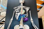
One case involved thoraco-omphalopagus twins joined at the chest and abdomen. Curione’s team 3D printed a multi-color anatomical model of the twins’ anatomy, which he called “the first of its kind for complexity in Italy.” Fabricated in transparent resin, the model offered a detailed view of the twins’ internal anatomy, including the rib cage, lungs, and cardiovascular system.
Attention then turned to the liver. The team built a digital reconstruction to simulate the optimal resection planes for the general separation and the hepatic splitting procedure. This was followed by a second multi-colour 3D printed model highlighting the organ’s vascularisation. These resources improved surgical planning, cutting operating time by 30%, and enabled a successful separation, with no major complications reported two years post-operation.
Dr. Davide Curione’s workflow for creating a 3D printed model of thoraco-omphalopagus twins using Mimics. Image via Frontiers in Physiology.
VR-enabled surgery enhances organ transplants
Materialise’s Mimics software can also be used in extended reality, allowing clinicians to interact more intuitively with 3D anatomical models and medical images. By using off-the-shelf virtual realityand augmented realityheadsets, healthcare professionals can more closely examine complex structures in an immersive environment.
Dr. David Sibřina is a Principal Researcher and Developer for the VRLab team at Prague’s Institute for Clinical and Experimental Medicine. He leads efforts to accelerate the clinical adoption of VR and AR in organ transplantation, surgical planning, and surgical guidance.
The former Forbes 30 Under 30 honouree explained that since 2016, IKEM’s 3D printing lab has focused on producing anatomical models to support liver and kidney donor programmes. His lab also fabricates 3D printed anatomical models of ventricles and aneurysms for clinical use.
However, Sibřina’s team recently became overwhelmed by high demand for physical models, with surgeons requesting additional 3D model processing options. This led Sibřina to create the IKEM VRLab, offering XR capabilities to help surgeons plan and conduct complex transplantation surgeries and resection procedures.
When turning to XR, Sibřina’s lab opted against adopting a ready-made software solution, instead developing its own from scratch. “The problem with some of the commercial solutions is capability and integration,” he explained. “The devices are incredibly difficult and expensive to integrate within medical systems, particularly in public hospitals.” He also pointed to user interface shortcomings and the lack of alignment with established medical protocols.
According to Sibřina, IKEM VRLab’s offering is a versatile and scalable VR system that is simple to use and customizable to different surgical disciplines. He described it as “Zoom for 3D planning,” enabling live virtual collaboration between medical professionals. It leverages joint CT and MRI acquisition models, developed with IKEM’s medical physicists and radiologists. Data from patient scans is converted into interactive digital reconstructions that can be leveraged for analysis and surgical planning.
IKEM VRLab also offers a virtual “Fitting Room,” which allows surgeons to assess whether a donor’s organ size matches the recipient’s body. A digital model is created for every deceased donor and live recipient’s body, enabling surgeons to perform the size allocation assessments.
Sibřina explained that this capability significantly reduces the number of recipients who would otherwise fail to be matched with a suitable donor. For example, 262 deceased liver donors have been processed for Fitting Room size allocations by IKEM VRLab. In 27 instances, the VR Fitting Room prevented potential recipients from being skipped in the waiting list based on standard biometrics, CT axis measurements, and BMI ratios.
Overall, 941 patient-specific visualizations have been performed using Sibřina’s technology. 285were for liver recipients, 311for liver donors, and 299for liver resection. Living liver donors account for 59cases, and split/reduced donors for 21.
A forum attendee using Materialise’s Mimics software in augmented reality. Photo via Materialise.
Personalized healthcare: 3D printing implants and surgical guides
Beyond surgical planning and 3D visualisation, Materialise Mimics software supports the design and production of patient-specific implants and surgical guides. The company conducts healthcare contract manufacturing at its Leuven HQ and medical 3D printing facility in Plymouth, Michigan.
Hospitals can design patient-specific medical devices in-house or collaborate with Materialise’s clinical engineers to develop custom components. Materialise then 3D prints these devices and ships them for clinical use. The Belgian company, headed by CEO Brigitte de Vet-Veithen, produces around 280,000 custom medical instruments each year, with 160,000 destined for the US market. These include personalised titanium cranio-maxillofacialimplants for facial reconstruction and colour-coded surgical guides.
Poole Hospital’s 3D specialists, Sian Campbell and Poppy Taylor-Crawford, shared how their team has adopted Materialise software to support complex CMF surgeries. Since acquiring the platform in 2022, they have developed digital workflows for planning and 3D printing patient-specific implants and surgical guides in 14 cases, particularly for facial reconstruction.
Campbell and Taylor-Crawford begin their workflow by importing patient CT and MRI data into Materialise’s Mimics Enlight CMF software. Automated tools handle initial segmentation, tumour resection planning, and the creation of cutting planes. For more complex cases involving fibula or scapula grafts, the team adapts these workflows to ensure precise alignment and fit of the bone graft within the defect.
Next, the surgical plan and anatomical data are transferred to Materialise 3-matic, where the team designs patient-specific resection guides, reconstruction plates, and implants. These designs are refined through close collaboration with surgeons, incorporating feedback to optimise geometry and fit. Virtual fit checks verify guide accuracy, while further analysis ensures compatibility with surgical instruments and operating constraints. Once validated, the guides and implants are 3D printed for surgery.
According to Campbell and Taylor-Crawford, these custom devices enable more accurate resections and implant placements. This improves surgical alignment and reduces theatre time by minimising intraoperative adjustments.
An example of the cranio-maxillofacial implants and surgical guides 3D printed by Materialise. Photo by 3D Printing Industry
Custom 3D printed implants are also fabricated at the Rizzoli Orthopaedic Institute in Bologna, Italy. Originally established as a motion analysis lab, the institute has expanded its expertise into surgical planning, biomechanical analysis, and now, personalized 3D printed implant design.
Dr. Alberto Leardini, Director of the Movement Analysis Laboratory, described his team’s patient-specific implant workflow. They combine CT and MRI scans to identify bone defects and tumour locations. Clinical engineers then use this data to build digital models and plan resections. They also design cutting guides and custom implants tailored to each patient’s anatomy.
These designs are refined in collaboration with surgeons before being outsourced to manufacturing partners for production. Importantly, this workflow internalizes design and planning phases. By hosting engineering and clinical teams together on-site, they aim to streamline decision-making and reduce lead times. Once the digital design is finalised, only the additive manufacturing step is outsourced, ensuring “zero distance” collaboration between teams.
Dr. Leardini emphasised that this approach improves clinical outcomes and promises economic benefits. While custom implants require more imaging and upfront planning, they reduce time in the operating theatre, shorten hospital stays, and minimise patient transfers.
After a full day of presentations inside the Irish College’s eighteenth-century chapel, the consensus was clear. 3D technology is not a niche capability reserved for high-end procedures, but a valuable tool enhancing everyday care for thousands of patients globally. From faster surgeries to cost savings and personalized treatments, hospitals are increasingly embedding 3D technology into routine care. Materialise’s software sits at the heart of this shift, enabling clinicians to deliver safer, smarter, and more efficient healthcare.
Take the 3DPI Reader Survey – shape the future of AM reporting in under 5 minutes.
Read all the 3D printing news from RAPID + TCT 2025
Subscribe to the 3D Printing Industry newsletter to keep up with the latest 3D printing news.You can also follow us on LinkedIn, and subscribe to the 3D Printing Industry Youtube channel to access more exclusive content.Featured image shows 3D printed anatomical models at Materialise HQ in Leuven. Photo by 3D Printing Industry.
#how #elevates #healthcare #insights #materialiseHow AM Elevates Healthcare: Insights from the Materialise 3D Printing in Hospitals Forum 2025
The cobbled streets and centuries-old university halls of Leuven recently served as a picturesque backdrop for the Materialise 3D Printing in Hospitals Forum 2025. Belgium’s Flemish Brabant capital hosted the annual meeting, which has become a key gathering for the medical 3D printing community since its launch in 2017.
This year, 140 international healthcare professionals convened for two days of talks, workshops, and lively discussion on how Materialise’s software enhances patient care. The Forum’s opening day, hosted at Leuven’s historic Irish College, featured 16 presentations by 18 healthcare clinicians and medical 3D printing experts.
While often described as the future of medicine, personalized healthcare has already become routine in many clinical settings. Speakers emphasized that 3D printing is no longer merely a “cool” innovation, but an essential tool that improves patient outcomes. “Personalized treatment is not just a vision for the future,” said Koen Peters, Executive Vice President Medical at Materialise. “It’s a reality we’re building together every day.”
During the forum, practitioners and clinical engineers demonstrated the critical role of Materialise’s software in medical workflows. Presentations highlighted value across a wide range of procedures, from brain tumour removal and organ transplantation to the separation of conjoined twins and maxillofacial implant surgeries. Several use cases demonstrated how 3D technology can reduce surgery times by up to four times, enhance patient recovery, and cut hospital costs by almost £6,000 per case.
140 visitors attended the Materialise 3D Printing in Hospitals Forum 2025. Photo via Materialise.
Digital simulation and 3D printing slash operating times
Headquartered a few miles outside Leuven’s medieval center, Materialise is a global leader in medical 3D printing and digital planning. Its Mimics software suite automatically converts CT and MRI scans into detailed 3D models. Clinicians use these tools to prepare for procedures, analyse anatomy, and create patient-specific models that enhance surgical planning.
So far, Materialise software has supported more than 500,000 patients and analysed over 6 million medical scans. One case that generated notable interest among the Forum’s attendees was that of Lisa Ferrie and Jiten Parmar from Leeds General Infirmary. The pair worked alongside Asim Sheikh, a Consultant Skullbase and Neurovascular Neurosurgeon, to conduct the UK’s first “coach door osteotomy” on Ruvimbo Kaviya, a 40-year-old nurse from Leeds.
This novel keyhole surgery successfully removed a brain tumor from Kaviya’s cavernous sinus, a hard-to-reach area behind the eyes. Most surgeries of this kind require large incisions and the removal of substantial skull sections, resulting in extended recovery time and the risk of postoperative complications. Such an approach would have presented serious risks for removing Kaviya’s tumor, which “was in a complex area surrounded by a lot of nerves,” explained Parmar, a Consultant in Maxillofacial Surgery.
Instead, the Leeds-based team uses a minimally invasive technique that requires only a 1.5 cm incision near the side of Ravimbo’s eyelid. A small section of skull bone was then shifted sideways and backward, much like a coach door sliding open, to create an access point for tumor removal. Following the procedure, Ravimbo recovered in a matter of days and was left with only a 6 mm scar at the incision point.
Materialise software played a vital role in facilitating this novel procedure. Ferrie is a Biomedical Engineer and 3D Planning Service Lead at Leeds Teaching Hospitals NHS Trust. She used mimics to convert medical scans into digital 3D models of Ravimbo’s skull. This allowed her team to conduct “virtual surgical planning” and practice the procedure in three dimensions, “to see if it’s going to work as we expect.”
Ferrie also fabricated life-sized, polyjet 3D printed anatomical models of Ravimbo’s skull for more hands-on surgical preparation. Sheikh and Parmar used these models in the hospital’s cadaver lab to rehearse the procedure until they were confident of a successful outcome. This 3D printing-enabled approach has since been repeated for additional cases, unlocking a new standard of care for patients with previously inoperable brain tumors.
The impact of 3D planning is striking. Average operating times fell from 8-12 hours to just 2-3 hours, and average patient discharge times dropped from 7-10 days to 2-3 days. These efficiencies translated into cost savings of £1,780 to £5,758 per case, while additional surgical capacity generated an average of £11,226 in income per operating list.
Jiten Parmarand Lisa Ferriepresenting at the Materialise 3D Printing in Hospitals Forum 2025. Photo via Materialise.
Dr. Davide Curione also discussed the value of virtual planning and 3D printing for surgical procedures. Based at Bambino Gesù Pediatric Hospital in Rome, the radiologist’s team conducts 3D modeling, visualization, simulation, and 3D printing.
One case involved thoraco-omphalopagus twins joined at the chest and abdomen. Curione’s team 3D printed a multi-color anatomical model of the twins’ anatomy, which he called “the first of its kind for complexity in Italy.” Fabricated in transparent resin, the model offered a detailed view of the twins’ internal anatomy, including the rib cage, lungs, and cardiovascular system.
Attention then turned to the liver. The team built a digital reconstruction to simulate the optimal resection planes for the general separation and the hepatic splitting procedure. This was followed by a second multi-colour 3D printed model highlighting the organ’s vascularisation. These resources improved surgical planning, cutting operating time by 30%, and enabled a successful separation, with no major complications reported two years post-operation.
Dr. Davide Curione’s workflow for creating a 3D printed model of thoraco-omphalopagus twins using Mimics. Image via Frontiers in Physiology.
VR-enabled surgery enhances organ transplants
Materialise’s Mimics software can also be used in extended reality, allowing clinicians to interact more intuitively with 3D anatomical models and medical images. By using off-the-shelf virtual realityand augmented realityheadsets, healthcare professionals can more closely examine complex structures in an immersive environment.
Dr. David Sibřina is a Principal Researcher and Developer for the VRLab team at Prague’s Institute for Clinical and Experimental Medicine. He leads efforts to accelerate the clinical adoption of VR and AR in organ transplantation, surgical planning, and surgical guidance.
The former Forbes 30 Under 30 honouree explained that since 2016, IKEM’s 3D printing lab has focused on producing anatomical models to support liver and kidney donor programmes. His lab also fabricates 3D printed anatomical models of ventricles and aneurysms for clinical use.
However, Sibřina’s team recently became overwhelmed by high demand for physical models, with surgeons requesting additional 3D model processing options. This led Sibřina to create the IKEM VRLab, offering XR capabilities to help surgeons plan and conduct complex transplantation surgeries and resection procedures.
When turning to XR, Sibřina’s lab opted against adopting a ready-made software solution, instead developing its own from scratch. “The problem with some of the commercial solutions is capability and integration,” he explained. “The devices are incredibly difficult and expensive to integrate within medical systems, particularly in public hospitals.” He also pointed to user interface shortcomings and the lack of alignment with established medical protocols.
According to Sibřina, IKEM VRLab’s offering is a versatile and scalable VR system that is simple to use and customizable to different surgical disciplines. He described it as “Zoom for 3D planning,” enabling live virtual collaboration between medical professionals. It leverages joint CT and MRI acquisition models, developed with IKEM’s medical physicists and radiologists. Data from patient scans is converted into interactive digital reconstructions that can be leveraged for analysis and surgical planning.
IKEM VRLab also offers a virtual “Fitting Room,” which allows surgeons to assess whether a donor’s organ size matches the recipient’s body. A digital model is created for every deceased donor and live recipient’s body, enabling surgeons to perform the size allocation assessments.
Sibřina explained that this capability significantly reduces the number of recipients who would otherwise fail to be matched with a suitable donor. For example, 262 deceased liver donors have been processed for Fitting Room size allocations by IKEM VRLab. In 27 instances, the VR Fitting Room prevented potential recipients from being skipped in the waiting list based on standard biometrics, CT axis measurements, and BMI ratios.
Overall, 941 patient-specific visualizations have been performed using Sibřina’s technology. 285were for liver recipients, 311for liver donors, and 299for liver resection. Living liver donors account for 59cases, and split/reduced donors for 21.
A forum attendee using Materialise’s Mimics software in augmented reality. Photo via Materialise.
Personalized healthcare: 3D printing implants and surgical guides
Beyond surgical planning and 3D visualisation, Materialise Mimics software supports the design and production of patient-specific implants and surgical guides. The company conducts healthcare contract manufacturing at its Leuven HQ and medical 3D printing facility in Plymouth, Michigan.
Hospitals can design patient-specific medical devices in-house or collaborate with Materialise’s clinical engineers to develop custom components. Materialise then 3D prints these devices and ships them for clinical use. The Belgian company, headed by CEO Brigitte de Vet-Veithen, produces around 280,000 custom medical instruments each year, with 160,000 destined for the US market. These include personalised titanium cranio-maxillofacialimplants for facial reconstruction and colour-coded surgical guides.
Poole Hospital’s 3D specialists, Sian Campbell and Poppy Taylor-Crawford, shared how their team has adopted Materialise software to support complex CMF surgeries. Since acquiring the platform in 2022, they have developed digital workflows for planning and 3D printing patient-specific implants and surgical guides in 14 cases, particularly for facial reconstruction.
Campbell and Taylor-Crawford begin their workflow by importing patient CT and MRI data into Materialise’s Mimics Enlight CMF software. Automated tools handle initial segmentation, tumour resection planning, and the creation of cutting planes. For more complex cases involving fibula or scapula grafts, the team adapts these workflows to ensure precise alignment and fit of the bone graft within the defect.
Next, the surgical plan and anatomical data are transferred to Materialise 3-matic, where the team designs patient-specific resection guides, reconstruction plates, and implants. These designs are refined through close collaboration with surgeons, incorporating feedback to optimise geometry and fit. Virtual fit checks verify guide accuracy, while further analysis ensures compatibility with surgical instruments and operating constraints. Once validated, the guides and implants are 3D printed for surgery.
According to Campbell and Taylor-Crawford, these custom devices enable more accurate resections and implant placements. This improves surgical alignment and reduces theatre time by minimising intraoperative adjustments.
An example of the cranio-maxillofacial implants and surgical guides 3D printed by Materialise. Photo by 3D Printing Industry
Custom 3D printed implants are also fabricated at the Rizzoli Orthopaedic Institute in Bologna, Italy. Originally established as a motion analysis lab, the institute has expanded its expertise into surgical planning, biomechanical analysis, and now, personalized 3D printed implant design.
Dr. Alberto Leardini, Director of the Movement Analysis Laboratory, described his team’s patient-specific implant workflow. They combine CT and MRI scans to identify bone defects and tumour locations. Clinical engineers then use this data to build digital models and plan resections. They also design cutting guides and custom implants tailored to each patient’s anatomy.
These designs are refined in collaboration with surgeons before being outsourced to manufacturing partners for production. Importantly, this workflow internalizes design and planning phases. By hosting engineering and clinical teams together on-site, they aim to streamline decision-making and reduce lead times. Once the digital design is finalised, only the additive manufacturing step is outsourced, ensuring “zero distance” collaboration between teams.
Dr. Leardini emphasised that this approach improves clinical outcomes and promises economic benefits. While custom implants require more imaging and upfront planning, they reduce time in the operating theatre, shorten hospital stays, and minimise patient transfers.
After a full day of presentations inside the Irish College’s eighteenth-century chapel, the consensus was clear. 3D technology is not a niche capability reserved for high-end procedures, but a valuable tool enhancing everyday care for thousands of patients globally. From faster surgeries to cost savings and personalized treatments, hospitals are increasingly embedding 3D technology into routine care. Materialise’s software sits at the heart of this shift, enabling clinicians to deliver safer, smarter, and more efficient healthcare.
Take the 3DPI Reader Survey – shape the future of AM reporting in under 5 minutes.
Read all the 3D printing news from RAPID + TCT 2025
Subscribe to the 3D Printing Industry newsletter to keep up with the latest 3D printing news.You can also follow us on LinkedIn, and subscribe to the 3D Printing Industry Youtube channel to access more exclusive content.Featured image shows 3D printed anatomical models at Materialise HQ in Leuven. Photo by 3D Printing Industry.
#how #elevates #healthcare #insights #materialise